CHOOSE YOUR CLINIC

Imaging techniques help doctors visualize internal body structures to diagnose various medical conditions. Some of the common ones are X-ray, CT scan, MRI, ultrasound, etc.
Diagnostic imaging refers to non-invasive medical tests that create images of the inside of the body to obtain clues about a medical condition. Based on this, doctors can diagnose diseases early and can guide targeted treatments. With high-invasive visualization, accurate diagnosis can be done.
In this article, we will talk about the various types of imaging techniques used in diagnosis.
Here are the key factors that contribute to choosing a technique.
| Factor | Description |
| Diagnostic need | Whether the issue is functional (metabolic) or structural (bone) |
| Safety concerns | Risk of ionizing radiation versus need for immediate diagnosis |
| Patient condition | Pregnancy (ultrasound), claustrophobia (MRI), etc. |
Table 1: Main factors behind selecting an imaging technique
Doctors adopt a range of techniques to diagnose various diseases. These include:
| Imaging technique | Imaging method | Best for |
| X-ray | Ionizing radiation | Bones, chest, teeth, etc. |
| MRI | Magnetic waves | Brain, soft tissue, ligaments, etc. |
| CT scan | Ionizing radiation | Urgent care, detailed 3D structure, etc. |
| Ultrasound | Sound waves | Abdomen, blood flow, pregnancy, etc. |
| PET scan | Radiotracers | Cancer, metabolic activities, etc. |
Table 2: X-ray vs CT scan vs MRI vs Ultrasound vs PET scan
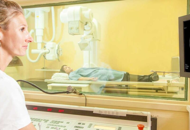
This is the oldest and one of the most common imaging techniques.
X-rays are quick and painless tests that produce images of the structures inside the body. Here, high-energy electromagnetic radiation is considered to produce static images of dense tissues like bones. The common types are chest X-rays, dental X-rays, bone density scans, etc.
When you enter the radiography room at DiagnostiX, the doctor will advise you to sit or lie on the bed. Here, the X-ray machine will capture images of your body’s internal structure.
You might be asked to move into several positions. The overall duration of the imaging technique is around 10 to 15 minutes.
An X-ray is used to diagnose various diseases. These include:
X-rays are essential for quick diagnostics, and this imaging technique stands for a high level of accuracy. However, ionizing radiation can contribute to increase cancer risk over many exposures.
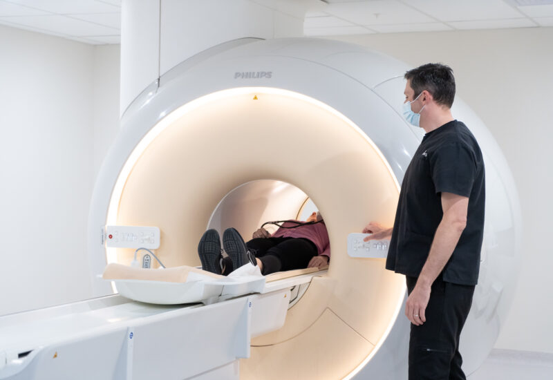
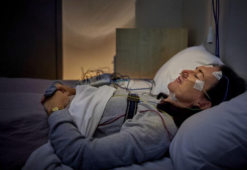
MRI creates detailed images of tissues and organs in the body.
An MRI is a non-invasive medical imaging test that creates detailed and 3D pictures of structures inside your body. How does MRI work? This technique uses magnetic field and radio waves.
Unlike X-rays, this test does not use any harmful ionizing radiation. The different types of MRI examinations are functional MRI, breast scans, cardiac MRI, etc.
During an MRI, you need to lie on a table that slides into the scanner. The machine produces tapping, loud knocking, or thumping noises.
Before you are placed on the MRI table, you need to remove all the metal objects. The overall duration is around 45 minutes to 1 hour.
MRI is suitable in the following scenarios:
There is no need for any ionizing radiation in an MRI. also, the procedure is a non-invasive one. However, the pitfalls of MRI scan are long scanning time, loud noise, and high cost.
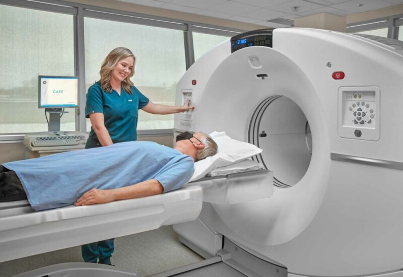
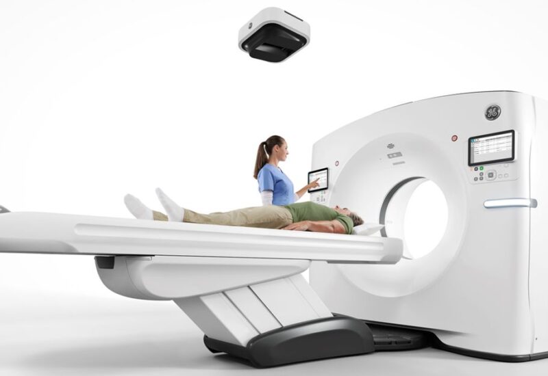
A CT (Computed Tomography) scan is a common imaging technique that uses the X-ray technique.
A CT scan is an imaging technique that considers a series of X-rays to create cross sections of bones, soft tissues, and blood vessels. A computer is used to create cross-sectional images. Compared to the conventional X-rays, a CT scan shows more detail.
As soon as you enter the scanning room, the doctor will advise you to lie on a table that slides into the scanner. This is followed by the rotation of the X-ray tube, which captures images. This procedure takes 10 to 15 minutes to complete.
A CT scan is appropriate in different scenarios, like:
CT scans are fast, which makes it effective in emergency situations. Also, this contributes to high detail imaging. Contrarily, there is a risk of radiation exposure.
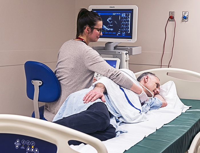
Ultrasound is a non-invasive imaging test that lets a healthcare provider to view inside of your body.
Ultrasound (also called sonography) uses high-frequency sound waves to create real-time pictures of internal organs. A device named a transducer emits the sounds and records echoes from the tissues.
While this procedure begins, a gel is applied to your skin. This is followed by passing a small probe against it. This is moved to capture images of the inside of your body. An ultrasound takes 30 minutes to 1 hour to complete.
You can consider an Ultrasound in Montreal because of the following instances:
The advantages of ultrasound are real-time imaging, wide accessibility, and no ionizing radiation. However, the drawbacks are limited penetration, limited deep imaging, and operator dependency.
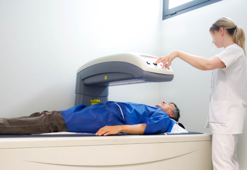
A PET scan is a functional imaging test that detects biochemical or metabolic changes.
PET (Positron Emission Tomography) scans refer to a type of imaging test where a little amount of radioactive tracer is used to measure and visualize metabolic activity in organs and body tissues. This tracer helps to detect diseased cells. This test highlights ‘hot spots’ where diseases are present.
During this test in DiagnostiX, the doctor injects a small amount of radiotracer into your arm. You need to sit or lie so that the tracer circulates through your body. A PET scan is a long process, and you have to be involved for 1.5 to 2 hours.
This imagining technique applies to:
The key positive sides of PET scans are excellent diagnostic accuracy and showing the functioning of organs. The downsides are radiation exposure, long duration, and expensiveness.
Imaging techniques contribute to disease identification so that treatment can be started quickly. The key imaging techniques are X-ray, CT scan, MRI, etc.
We are a leading clinic for imaging techniques in Montreal. Visit our clinic to and get an expert consultation. This lets you understand the one you need. Get it done swiftly.
X-rays are generally avoided during pregnancy. This is because the imaging test prevents exposing the rapidly developing fetus to ionizing radiation. This increases the risk of birth defects. Inform your doctor if you are pregnant.
You can eat, drink, and take medicines normally before an MRI scan. Some specialized scans require fasting for 4 to 6 hours. Avoid wearing clothing with zippers or metal snaps when you go for an MRI.
PET scans or CT scans are perfect to detect cancer. This is because of the combination of structural (CT) and metabolic (PET) imaging to identify active tumor cells. Well, MRI is used for specific organs.
The preparation for an ultrasound depends on the area that is to be scanned. In the case of abdomen, avoid eating or drinking for 8 to 12 hours to avoid intestinal gas. For pelvic sonography, drink 4 to 6 glasses of water an hour prior to the appointment. In regards to kidney, there is a need for a combination of fasting and a full bladder.
In short, yes. You need a referral (also called a requisition) from a qualified medical practitioner like a specialist, a nurse practitioner, or a family doctor. This is a common requirement for both public health coverage and private clinics.