CHOISISSEZ VOTRE CLINIQUE

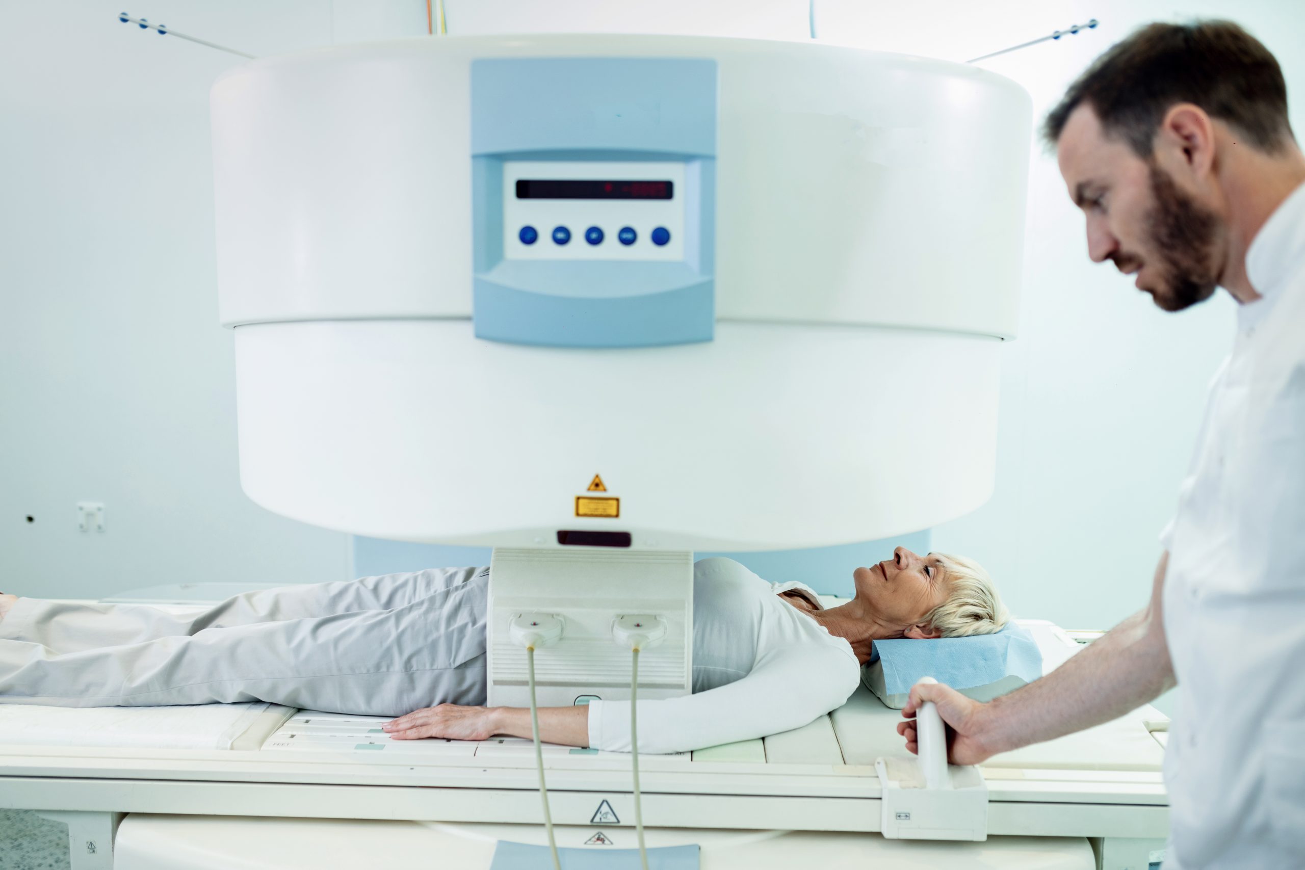
MRI is a process of diagnostic imaging, and it can diagnose the brain, spine, musculoskeletal health, abdomen, pelvis, and breasts. Before getting an MRI, you need to follow some rules, such as removing all metallic objects from your body, not eating before an abdominal MRI, and so on.
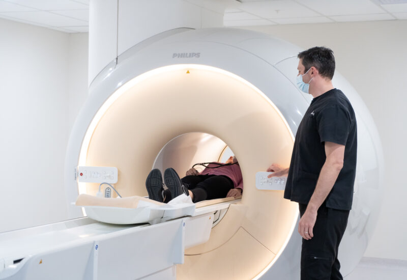
MRI or Magnetic Resonance Imaging is a non-invasive medical test that creates highly detailed and cross-sectional images inside your body. MRI is completely safe compared to X-rays or CT scans, as it does not use any ionizing radiation.
MRI technology works using magnetic fields, radio waves, and energy signals.
How it actually works:
People are shifting towards MRI in Montreal because it is a comparatively safer testing procedure than CT scans and X-rays.
Why it is considered safe:
When booking a private MRI in Quebec, you will often choose between a 1.5 Tesla (1.5T) and a 3 Tesla (3T) scanner. The "T" stands for Tesla—the universal unit of measurement for magnetic field strength.
While a 3T magnet is exactly twice as powerful as a standard 1.5T magnet, "more powerful" does not automatically mean "better" for every medical situation. The right scanner depends entirely on the specific organ being examined, your medical history, and the presence of any internal medical hardware.
| Feature | 1.5T MRI Scanners | 3T MRI Scanners |
|---|---|---|
| Magnetic Field Strength | Standard clinical baseline (~30,000 times stronger than Earth's magnetic field). | High-field clinical strength (~60,000 times stronger than Earth's magnetic field). |
| Signal-to-Noise Ratio (SNR) | Standard baseline; provides clear, highly reliable imaging for routine diagnostic paths. | Doubled SNR; allows for ultra-high spatial resolution and microscopic structural tracking. |
| Average Scan Speed | Standard exam durations (typically 20–45 minutes depending on the target protocol). | Up to 30–50% faster scan times, significantly reducing the window for accidental patient motion. |
| Best Used For | Routine spine work, abdominal imaging, and patients with compatible metallic implants. | Advanced neurology (brain mapping, stroke, MS), micro-MSK joint tears, and prostate imaging. |
| Claustrophobia and Control | Available in wide-bore configurations, but can sometimes feel tighter depending on the older scanner model. | Often built with wider, shorter bores and faster scan speeds, significantly reducing claustrophobic anxiety. |
|
Susceptibility Artifacts |
Low. Highly stable around surgical clips, pins, or screws; causes less image distortion near metal. | High. Highly sensitive to metal; minor magnetic implants can cause large blind spots (artifacts) on the scan. |
Table: 1.5T vs. 3T
A common misconception in health blogging is that 3T scanners inherently have wider tunnels (bores) than 1.5T systems. This is technically incorrect.
Bore architecture—whether a machine is a standard 60cm tunnel or a spacious 70cm "wide-bore"—is entirely an engineering choice made by manufacturers (like Siemens, GE, or Philips), completely independent of the magnet's power.
Many modern 1.5T scanners feature 70cm wide-bores specifically designed to alleviate patient anxiety.
Conversely, some older 3T systems feature narrower tunnels to sustain their intense magnetic fields.
At modern private clinics, claustrophobia is mitigated not by the Tesla rating, but by choosing short-bore, wide-bore machine designs coupled with rapid scan sequences that minimize the time you spend inside the machine.
When 3T is Imperative: Your referring physician will typically request a 3T scan for complex neurological or minute orthopedic evaluations. The ultra-high resolution is vital for mapping tiny vascular pathways in the brain, identifying early-stage plaques in Multiple Sclerosis, detecting micro-tears in the wrist or ankle cartilage, or performing multi-parametric prostate imaging.
When 1.5T is Safest: A 1.5T system remains the gold standard for patients with structural medical implants (such as older orthopedic screws, artificial joints, or certain pacemakers). Because higher magnetic fields amplify distortion, a 1.5T scanner minimizes "magnetic artifacts," allowing radiologists to see the surrounding tissues clearly without structural interference or unnecessary safety risks.
While the MRI cost for a full-body scan ranges between $1500 and $3650 CAD, different types of MRI scans target different anatomical zones in your body, depending on what you need.
Brain MRI is a painless imaging test that generates highly detailed, cross-sectional pictures of your brain and its intricate structures.
This diagnostic imaging procedure is used to create detailed images of bones, joints, muscles, ligaments, and tendons. To assess any tear, inflammation, or sports injuries, getting an MRI done is a smarter choice to avoid any unexpected health issues.
This is done to diagnose, monitor, or rule out different types of health conditions in the abdomen and pelvis.
Private breast MRI services are highly sought after across Quebec due to the critical nature of early detection and the desire to completely bypass public hospital waitlists.
Important Legal Notice for Quebec Patients: In strict compliance with the Collège des médecins du Québec, all diagnostic imaging exams—including those performed at private clinics—legally require a valid medical prescription (requisition) signed by a licensed healthcare practitioner. If you do not currently have a referral, DiagnostiX can seamlessly coordinate an expedited consultation with a partner telemedicine network or a licensed general practitioner to evaluate your clinical eligibility and issue the necessary ordonnance médicale.
High-Risk Cancer Screening Supplement: For patients with a dense breast tissue profile, a confirmed BRCA1 or BRCA2 gene mutation, or a strong family history of breast cancer, an MRI is utilized as a vital secondary screening tool alongside standard digital mammography.
Evaluating Breast Implant Integrity: It stands as the gold-standard diagnostic tool for checking both silicone and saline breast implants, providing unmatched structural clarity to identify subtle intracapsular or extracapsular ruptures that ultrasounds might miss.
Staging and Post-Treatment Monitoring: For patients with a recent diagnosis, it precisely maps tumor boundaries and scans for multi-focal disease prior to surgical planning.
Prone Position Layout: Unlike standard supine (face-up) scans, you will lie face down on a specialized, ergonomically padded table. Your breasts will rest comfortably inside a dedicated breast coil—a specialized signal antenna engineered to capture high-resolution tissue data without compressing the anatomy.
Intravenous Contrast (Gadolinium): To accurately differentiate between benign cysts and highly vascularized abnormal tissues, a breast MRI almost always requires an injection of a gadolinium-based contrast agent through an IV line in your arm midway through the scan.
Many targeted MRI scans—including detailed neurological, vascular, and breast imaging—require an intravenous injection of a Gadolinium-Based Contrast Agent (GBCA). Gadolinium is a paramagnetic metal that temporarily alters how water molecules inside your body react to magnetic forces, highlighting blood vessels, tumor margins, and focal tissue inflammation with pristine diagnostic clarity.
While the phrase "chemical contrast" can cause patient anxiety, modern MRI contrast protocols are exceptionally safe and highly regulated.
Historically, medical concerns surrounding MRI contrast were linked to older, "linear" chelate designs. If the contrast remained in the system of a patient with severe kidney failure for an extended period, these older compounds could break down, allowing free gadolinium to deposit in tissues and cause a rare, severe condition called Nephrogenic Systemic Fibrosis (NSF).
Today, premier private clinics across Canada have completely eliminated this hazard by adhering to updated safety frameworks set by the Canadian Association of Radiologists (CAR) and Health Canada:
Because gadolinium contrast is processed and cleared entirely by your renal system, your kidneys are responsible for flushing the agent from your bloodstream.
To guarantee patient safety, private clinics screen kidney health using an eGFR (Estimated Glomerular Filtration Rate) blood test result—typically required to be taken within 30 to 90 days of the scan—for individuals who meet any of the following criteria:
What the Metrics Mean: An eGFR score of 30 mL/min/1.73m² or higher is considered a safe clearance baseline for a standard macrocyclic contrast dose. If an outpatient's eGFR falls below 30, a radiologist will personally conduct a strict risk-benefit audit, consult your referring physician, and evaluate whether a non-contrast sequence can adequately capture the diagnostic data.
For any patient with baseline kidney health, over 95% of the administered contrast is naturally excreted through urine within 24 hours.
The clinical preparation for clearing contrast is straightforward: expand your fluid intake. Drinking an extra two to three glasses of water immediately following your procedure accelerates renal filtration, allowing your body to flush out remaining traces of the agent seamlessly.
Before the MRI scan, there are some rules and regulations you need to follow to ensure safety.
If you are scheduled for an MRI of your abdomen or pelvis, you will be instructed to fast—consuming no solid foods or liquids (except small sips of water)—for 4 to 6 hours before your appointment.
This is not a random rule; it is a strict clinical necessity required to stop internal organ movement and structural shifts that can completely ruin the diagnostic quality of the scan.
Eating or drinking before the scan triggers two distinct physiological responses that blur the final images:
The moment food or liquid enters your stomach, your digestive system activates peristalsis—the involuntary, wave-like muscular contractions that ripple through the stomach and intestines to push contents forward.
Because an MRI takes several minutes per sequence to capture high-resolution images, even microscopic movement inside the body acts like a camera shaking during a long-exposure photo. Fasting ensures the gastrointestinal tract is entirely at rest, preventing motion artifacts (imaging blur) and allowing the radiologist to clearly view static organs like the pancreas, liver, and kidneys.
To accurately evaluate the gallbladder and bile ducts—especially during specialized protocols like an MRCP (Magnetic Resonance Cholangiopancreatography)—the gallbladder must be fully distended (expanded and filled with bile).
When you ingest food, particularly fats, your small intestine releases hormones that trigger gallbladder contraction. As the gallbladder contracts to pump bile into the digestive tract, it deflates and collapses. A collapsed gallbladder makes it nearly impossible for a radiologist to detect tiny pathologies, such as hidden gallstones, early-stage polyps, or subtle thickening of the gallbladder wall.
Clinical Takeaway: Arriving at the clinic in a true fasting state ensures your internal organs are perfectly still and fully distended. This guarantees the highest possible image clarity on the first attempt, preventing the need for frustrating re-scans or inconclusive results.
If you are prescribed an MRI test, keep the following things in mind for a better experience.
MRI is an advanced imaging procedure for the screening of your inner organs, like the brain, spine, pelvic area, and abdomen, and so on, so that the doctors can detect and diagnose any abnormal condition at an early stage and begin the necessary treatment right away to avoid any type of fatal consequences.
If you are planning to get an MRI, book an appointment at DiagnostiX for a smooth procedure. You can also call us at 438-858-8611 for further details.
No, you cannot wear regular clothes unless they are 100% metal-free. At DiagnostiX, we provide a medical gown or clinic-provided, metal-free cotton scrubs.
The "T" stands for Tesla, which is the unit of measurement for magnetic field strength. A 3T MRI machine produces a magnetic field that is twice as strong as a conventional 1.5T scanner. This increased strength generates a much higher signal-to-noise ratio, which translates directly into ultra-high-resolution, incredibly detailed images
Most modern body piercings are made from non-ferrous metals like high-grade titanium or surgical stainless steel, but all external jewelry must still be removed before your scan to prevent image distortion.
A: A standard MRI examination typically takes between 15 and 45 minutes per body part, depending on the complexity of the area and whether IV contrast is required. A definitive diagnostic report is compiled and delivered directly to your referring healthcare provider within 24 to 48 business hours.